Back Pain in the Horse: Common Conditions & Diagnosis
- Jul 5, 2025
- 4 min read
Updated: Jul 11, 2025
Explore the causes of back pain in the horse, including commonly diagnosed conditions like kissing spines, arthritis, and soft tissue problems. Learn how clinical assessment and imaging lead to accurate diagnosis.

Introduction
Back pain in the horse is a common yet often under-recognised issue affecting both comfort and performance.
The thoracolumbar spine, the combination of the thoracic and lumbar spine, plays a crucial role in transferring force, supporting the rider and stabilising the body during movement. Lesions in this region can lead to subtle performance changes, postural compensation, or overt discomfort under saddle.
A structured diagnostic approach, combining physical examination, objective gait analysis, and advanced imaging, is essential to accurately identify the cause of thoracolumbar pain and guide targeted treatment.
Common Thoracolumbar Spine Lesions in Horses
1. Dorsal Spinous Process Impingement (“Kissing Spines”)


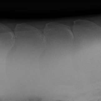


Varying degrees of impinging spinous process from mild to severe.
Characterised by the close approximation, contact, or overlapping of adjacent dorsal spinous processes, commonly between T14 and T18. Associated pathology may include reactive sclerosis, lysis, or interspinous ligament entrapment. Diagnosis is via x-ray and active lesions via scintigraphy. Quite often
Clinical signs:
Resistance to saddling or mounting
Discomfort on palpation, tense muscle
Hollowing or bracing of the back under saddle
Reduced performance or altered back use
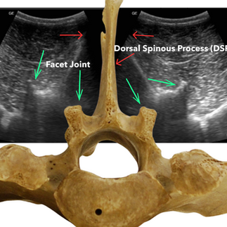
2. Facet Joint Osteoarthritis (arthritis of the joints)

Degenerative changes affecting the synovial intervertebral (facet) joints can be frequently seen in the caudal thoracic and lumbar regions.
This area can often be overlooked due to the more technical ultrasonography skils orequired to assess this region. This area is unable to be seen on regular field x-rays due to the tissue thickness.
Clinical signs:
Generalised back stiffness
Reluctance in lateral work or collection
Pain on back extension
Behavioural issues under saddle
3. Supraspinous & Interspinous Ligament Desmitis
These ligaments help stabilise the dorsal spinous processes. Repetitive strain, ill-fitting tack, or trauma can lead to fibre disruption and chronic pain. Diagnosis involves the use of ultrasonography.
Clinical signs:
Localised pain on palpation
Reduced back flexibility
Sensitivity when grooming or saddling
4. Epaxial Muscle Strain or Atrophy


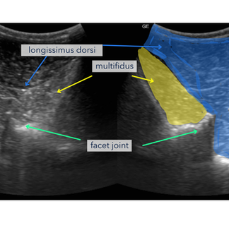

The longissimus dorsi and multifidus are essential for stabilising and supporting the spine. Injury or chronic overload leads to reactive tension, loss of symmetry, or postural compromise. Diganosis is via ultrasonographic assessment, muscle scoring and palpation.
Clinical signs:
Poor topline or visible muscle wasting
Asymmetrical movement
Increased muscle tone or fasciculations
Discomfort during spinal mobilisation
5. Vertebral Body or Spinous Process Fractures

Less commonly but important in cases of acute trauma. Fractures can involve the vertebral body, joints or dorsal spinous processes. Diagnostics in regards to DSP fractures can be straight forward however for deeper tissues such as the vertebral bodies, scinitigraphy is the modality of choice.
Clinical signs:
Sudden-onset severe pain
Localised heat, swelling, or neurological signs
Gait abnormalities or instability
Approach to Back Pain in the Horse
A systematic approach is essential when working up back pain in the horse. A detailed history (behavioural and work) in combination with a detailed physical examination and chiropractic assessment by a veterinary horse chiropractor (or veterinary physio) is key.
Combining these assessments with a gait assessment to rule out a primary or contributing lameness is also important. If there is a lameness present this needs to be identified with a nerve block assessment, sometimes lamenesses do not block to the limbs and this is where the contribution of higher up structures like the neck, back and pelvis need to be considered.
Even when a lameness is present it doesn't necessarily mean there are no other problems in the spinal region. This is where combining the history, gait assessment and chiropractic exam become essential.
Diagnostics
When trying to diagnose back pain in the horse, the main imaging modalities available include:
X-rays
Ultrasound
Scintigraphy.

X-rays: Due to the size of horses back x-rays are limited (unless in a hospital setting with very high powered units) to assessing the dorsal spinous processes and in some locations (such as the thoracic spine) the facet joints. Visualising the lumbar facet joints is difficult and images are often non-diagnostic.
X-rays assess for bone change such as remodelling and changes at the bony surface where ligaments attach. As such, combining x-rays with ultrasonographic examination is very useful.
Ultrasound:

Ultrasound is very useful to penetrate to deeper tissues of the horse's back. We use it to identify bone changes at the level of the joints (arthritis) and for a assessing soft tissues (ligaments & muscle).
Ultrasound guided treatments ensure that specific structures are targeted to have the greatest effect possible.
Scintigraphy: Highlights areas of active bone turnover and helps detect deep bone related discomfort, arthritis and potential fractures.
Conclusion
Back pain in the horse is frequently linked to dysfunction of the thoracolumbar spine. While signs may be subtle, early recognition and accurate diagnosis are essential to prevent secondary compensation and ensure long-term soundness. A structured clinical approach, incorporating physical exam, gait analysis, and imaging, allows for targeted intervention and measurable improvements in both comfort and performance.
Need Support with a Suspected Spinal Case?
At Core Equine, we provide comprehensive spinal diagnostics tailored to sport and performance horses, including:
Veterinary chiropractic evaluation
SLEIP-based objective gait analysis
High-resolution ultrasound of the horse's back
Image-guided therapies and rehab plans
Whether you're looking for a trusted lameness vet in Melbourne or a comprehensive assessment of horse lameness, we’re here to help.


Comments