


Client Journey: Mack
- Anushka von Oppen
- Feb 21
- 3 min read
This month we are going to take a look at a client journey.
If you would like to understand the data prior to reading this blog, you can have a look at my SLEIP explainer here.
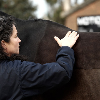
We’ve had the privilege of caring for Mack and his dedicated owner for almost a year. Initially, Mack began with routine gait assessments and chiropractic sessions every 4–6 weeks. During this time, a significant lameness issue emerged, but through early detection, targeted joint management, and a committed rehabilitation plan, we were able to not only address the problem but also enhance his overall strength and suppleness. Today, Mack is physically stronger and more supple than before we started seeing him, meaning we have increased the time even further between assessments. It is a real testament to proactive care and dedicated teamwork.

Here is a comparison of Mack’s gait between July and September. The sudden onset of right hind (RH) lameness is highlighted by the red bars on the right side of the image.
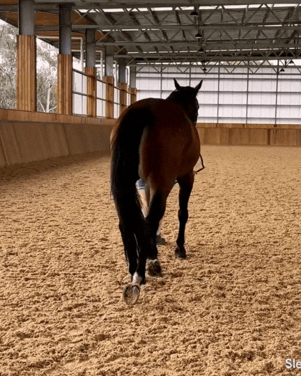
To investigate further, we performed a series of nerve blocks, progressing up the limb. However, even after the third block (tibial peroneal), which desensitises structures below the hock, there was no significant improvement (see image below).
Given the presence of mild joint filling in the right stifle, we proceeded with an ultrasound, revealing chronic changes within the joint.
What stood out in Mack’s case—similar to many others we see—was his relative historical soundness despite these underlying issues. This gave us confidence that, with the right management, we could not only restore his soundness but also ensure he remains comfortable and continues to be enjoyed by his dedicated owner.
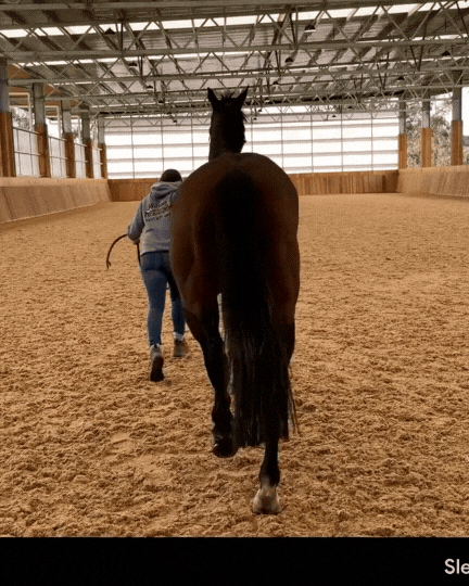


Video on the left: pre-block trot out; Image on the right: documenting the nerve blocking of the distal limb without significant improvement to the lameness.

Given the versatility of ultrasonography in assessing both bone surfaces and soft tissues, we chose to use it as our primary imaging modality.
The ultrasound revealed bilateral joint filling in the left and right medial femorotibial joints (FTJ), along with bone remodeling on the inner aspect of the femur and signs of joint-related inflammation.

Ultrasound also revealed evidence of meniscal fiber disruption. The meniscus is a crucial ligament that acts as a shock absorber, providing a cushioned and stable surface for the femur to articulate smoothly over the relatively flat tibia(see anatomy here.)


Following medication of the joint Mack's lameness has significantly improved.
After diagnosing inflammation and arthritis in both stifles, we initiated targeted management of the medial femorotibial joints. Given the sudden onset of lameness and the degree of inflammation present, initial therapy included a combination of corticosteroids and hyaluronic acid to reduce inflammation and support joint function.
As part of a longer-term approach to soft tissue repair, we later treated the joint with Pro-Stride® APS—a regenerative therapy containing a concentrated solution of white blood cells, platelets, growth factors, and anti-inflammatory cytokines. This advanced treatment helps reduce inflammation while promoting soft tissue healing.
Alongside medical intervention, Mack’s owner implemented a structured rehabilitation program implementing exercises from the EqActive app, which played a crucial role in his recovery. She was diligent in completing stretches, strengthening exercises, and incorporating the Equiband® system to enhance hindquarter strength and stability. I firmly believe that without her commitment to physiotherapy-based rehabilitation, we would not have achieved such positive results.
While corticosteroids provided significant pain relief, Mack’s overall improvement highlights the importance of a whole-horse approach—where medical treatment, rehabilitation, and owner dedication work together for optimal long-term outcomes.

Comentarios